Results
2 results found
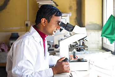
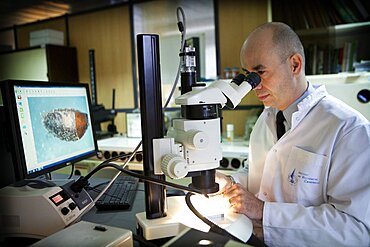
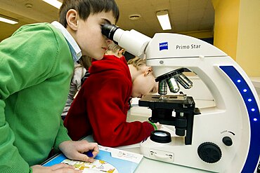
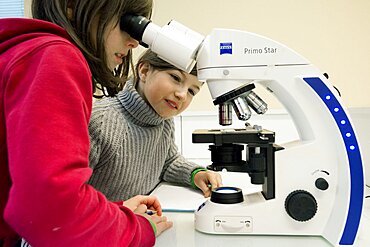
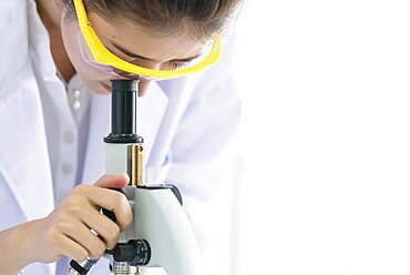
A lab technician working in a laboratory in a small hospital in Nepal looks into a microscope, Jiri, Solu Khumbu, Nepal, Asia
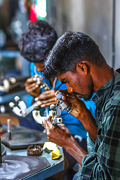
Diamond polishing workshop in a village near Dediapada in Narmada district, Gujarat, India, Asia
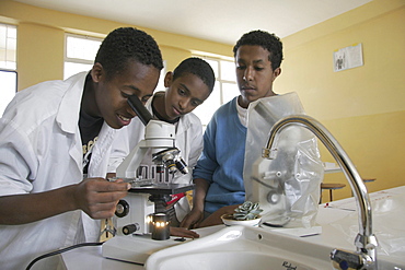
Ethiopia science class for 10th grade at the lazarist catholic high school addis ababa

Ethiopia science class for 10th grade at the lazarist catholic high school addis ababa
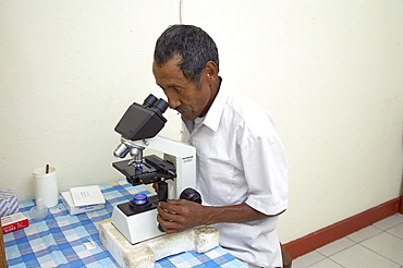
East timor. Examining blood sample to test for malaria, aileu
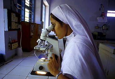
Technology nurse microscope, damien leprosy center, trichur, india
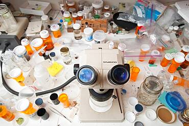
Lab facility with desktop of samples bottles, microscope, pill bottles, petri dishes
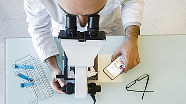
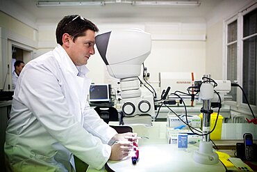
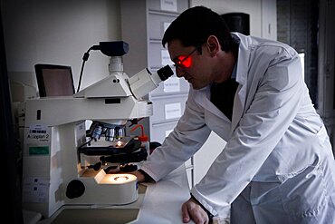
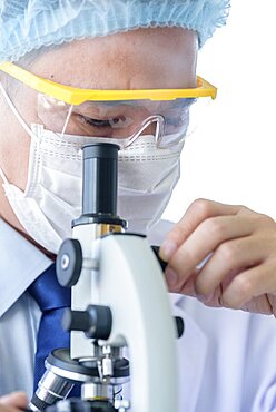
Mature scientist male in his 50s wearing a lab coat looking through a microscope in a laboratory. Basque Country, Spain, Europe.
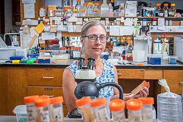
University of Georgia researcher, Peggy Ozias-Akins;, sitting in front of microscope in her lab researching molecular genetics of peanut plants, Tifton, Georgia.
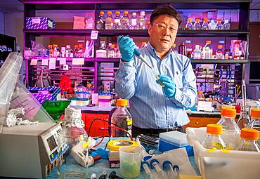
Male scientist in lab using pipette surrounded by lab equipment.
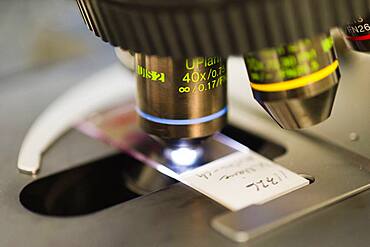
Microscope
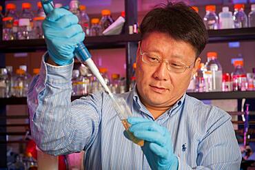
Male scientist in lab using pipette.
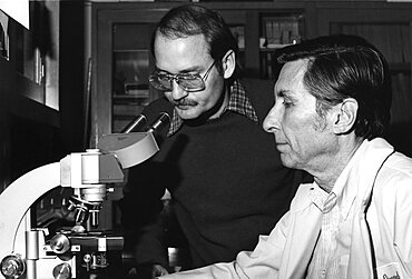
Discovering Legionnaires' Disease, 1977
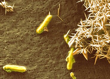
Alkali-loving extremophile bacteria, SEM
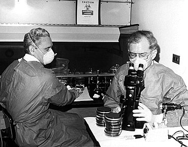
Discovering Legionnaires' Disease, 1978
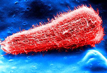
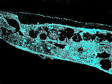
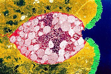
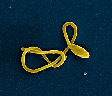
Miracidium larva of the liver fluke (fasciola hepatica) viewed in SEM.
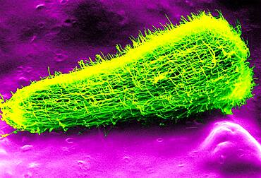
Miracidium larva of the liver fluke (fasciola hepatica) viewed in SEM.

Birch pollen, sem
Cowpox virus, used for the preparaton of a smallpox vaccine (TEM). Electron micrograph of a Vaccinia Virus. Vaccinia virus is normally confined to cattle, but is conveyed to humans through vaccination
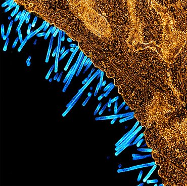
Syphilis bacterium. Treponema pallidum subsp pallidum on cultures of cotton tail rabbit epithelium cells Sf1Ep
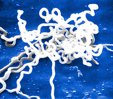
Bacterium responsible for syphilis. Electron micrograph of Treponema pallidum on cultures of cotton-tail rabbit epithelium cells (Sf1Ep).Treponema pallidum is the causative agent of syphilis. In the United States, over 35,600 cases of syphilis were repor

Diatom, Sem
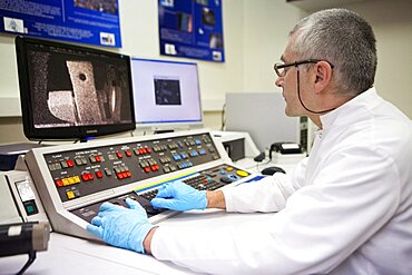
Crime Scene Investigation
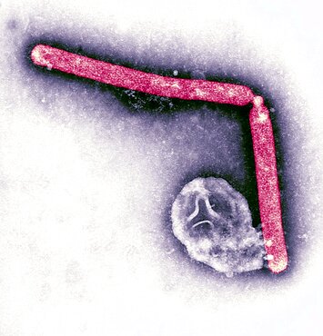
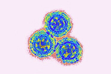
Avian Influenza Virus

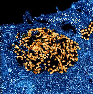
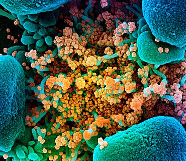
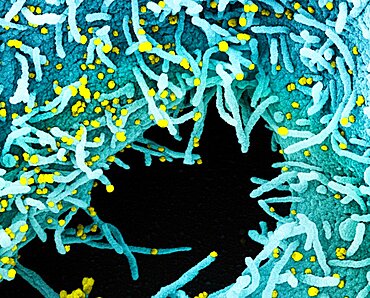
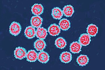
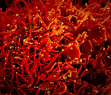
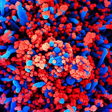
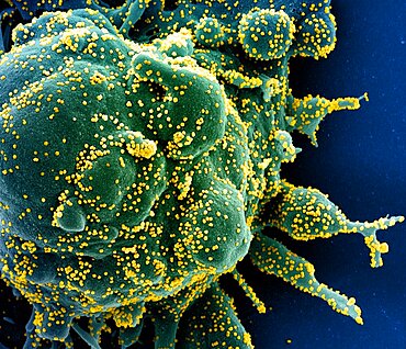
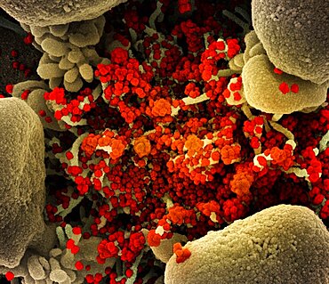
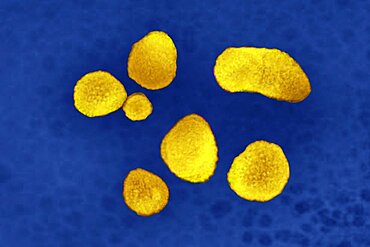
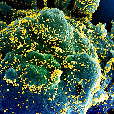
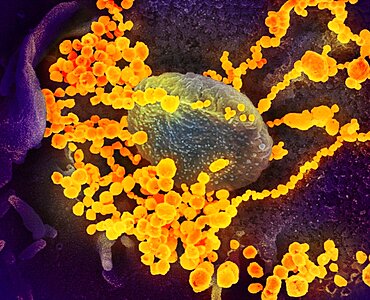
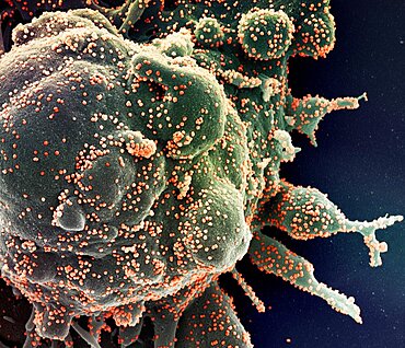
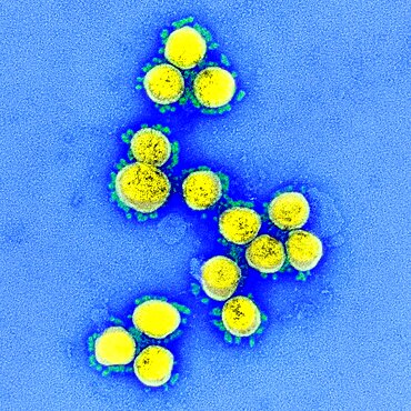
Novel coronavirus sars-cov-2
Crime Scene Investigation

Diatom, Sem
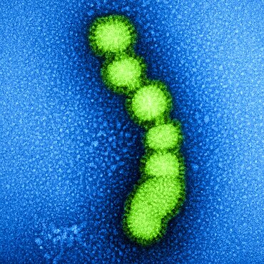
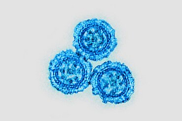
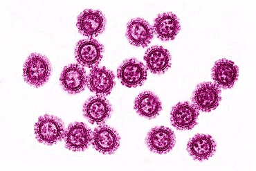
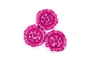
Swine flu strain virus particles

Micrograph of methicillin-resistant staphylococcus aureus (mrsa)

Diatom, Sem

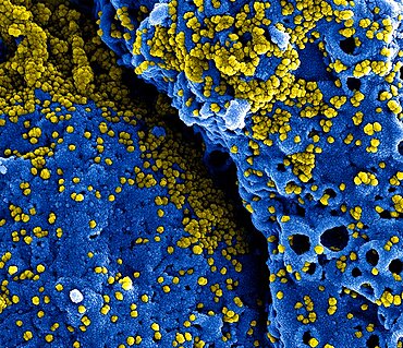
Novel coronavirus sars-cov-2

Diatom, Sem

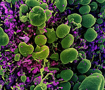
Novel coronavirus sars-cov-2

Diatom, Sem
Crime Scene Investigation
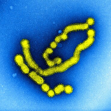
Swine flu strain virus particles
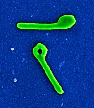
Ebola virus nucleocapsids and virus particles
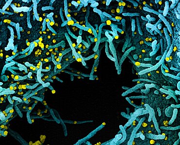
Novel coronavirus sars-cov-2
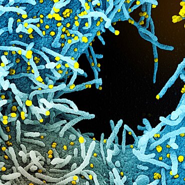
Novel coronavirus sars-cov-2
Crime Scene Investigation
Novel coronavirus sars-cov-2
Novel coronavirus sars-cov-2

Kid Campus Workshop
Goblet Cell, Tem
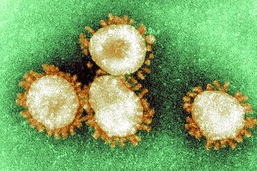
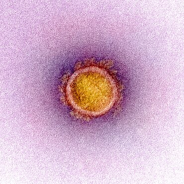
Coronavirus, Tem
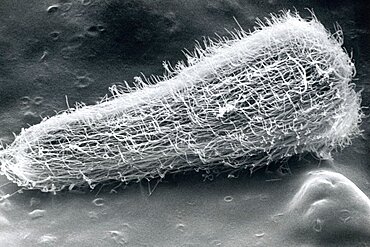
Sheep Liver Fluke Larva
Coronavirus
Coronavirus
Coronavirus
Coronavirus
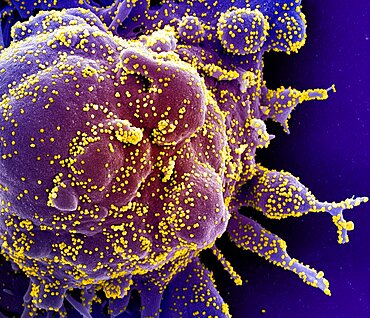
Novel coronavirus sars-cov-2
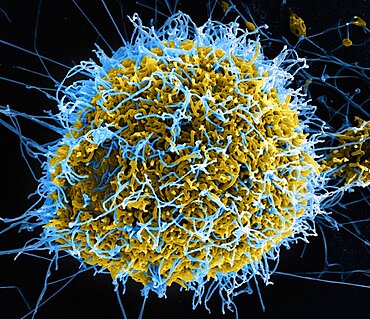
Ebola virus particles
Novel coronavirus sars-cov-2

Novel coronavirus sars-cov-2
Novel coronavirus sars-cov-2
Two ebola virus particles
Kid Campus Workshop
Kid Campus Workshop
Ebola virus particle
Swine flu virus particles
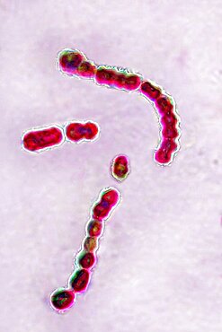
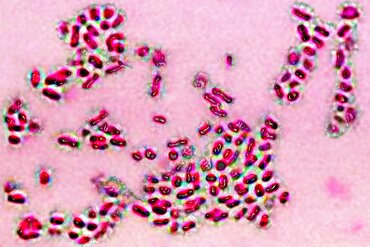
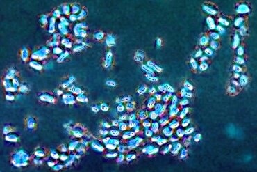
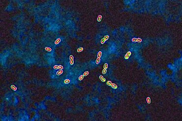
Streptococcus pyogenes
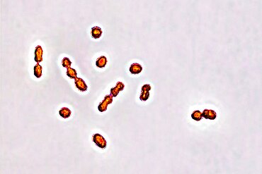
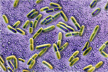
Prevotella bacteria
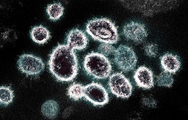
Novel coronavirus sars-cov-2
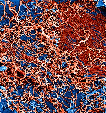
Ebola virus particles
Prevotella bacteria
Novel coronavirus sars-cov-2
Novel coronavirus sars-cov-2
Streptococcus agalactiae
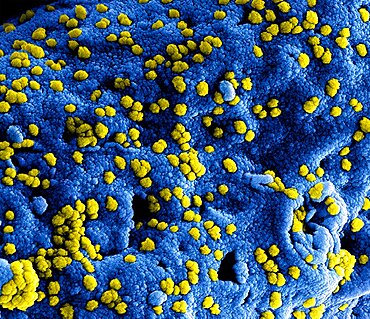
Mers coronavirus particles

Ebola virus

Pneumococcal bacteria
Pneumococcal bacteria
Prevotella bacteria
Asian young man student scientist researching and learning in a laboratory.
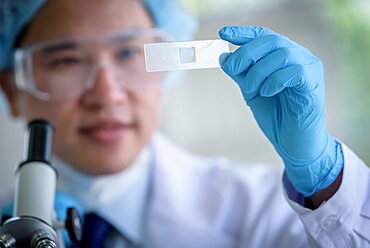
Asian man scientist researching and learning in a laboratory.

Asian young girl student scientist researching and learning in a laboratory.

Asian young girl student scientist researching and learning in a laboratory.

Asian senior scientist researching and learning in a laboratory.
Asian young student scientist researching with a microscope in a laboratory.

Asian senior scientist male researching and learning in a laboratory.

Asian young girl student scientist researching and learning in a laboratory.
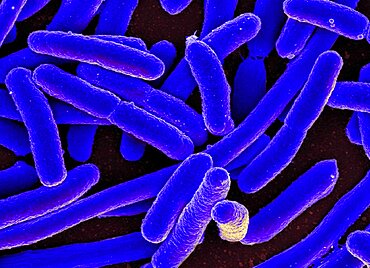
Colorized scanning electron micrograph of Escherichia coli, grown in culture and adhered to a cover slip.
Novel coronavirus sars-cov-2
Novel coronavirus sars-cov-2
Novel coronavirus sars-cov-2
Mers coronavirus particles
Coronavirus
Novel coronavirus sars-cov-2
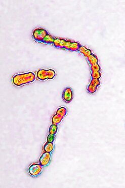
Streptococcus pyogenes
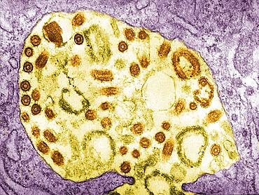
Electron micrograph of the Marburg virus. Marburg virus, first recognized in 1967, causes a sever type of hemorrhagic fever, which affects humans, as well as non-human primates.
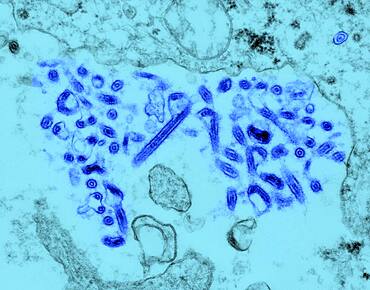
Ebola Virus viewed under TEM. Ebola virus is a RNA virus. It is responsible for severe hemorrhagic fever.
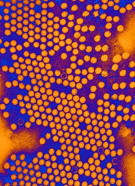
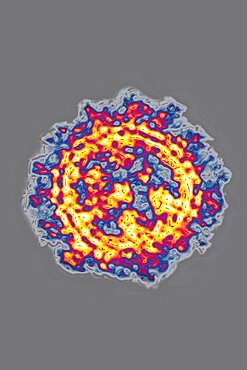
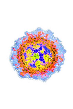
Electron micrograph of the poliovirus. Poliovirus is a species of Enterovirus, which is a genus in the family of Picornaviridae, and is an RNA virus.

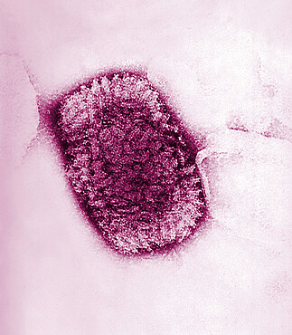
Measles virus (of large size) and SV40 virus (of small size), TEM. This electron micrograph reveals both a paramyxovirus measles virus, and virions of the polyomavirus, simian virus SV40 (smaller circles)
Novel coronavirus sars-cov-2

Coronavirus
Coronavirus
Coronavirus

Pneumococcal bacteria
Escherichia Coli, SEM
Novel coronavirus sars-cov-2